Most of the conversation around clavicular development treats the topic as a binary: either your growth plates are open and you can still add shoulder width, or they're closed and you can't. The published data is more interesting than that. The medial clavicular epiphysis is not only the last growth plate in the body to fuse — it fuses across a range of ages wide enough that two healthy adults born in the same year can be in entirely different biological windows at 24. Understanding that range, and the longitudinal growth numbers that go with it, is the foundation for any serious research discussion about frame optimization.
The Numbers That Actually Matter
Hughes and colleagues, in a longitudinal study published in 2020, tracked clavicular length in a cohort of healthy adolescents and young adults from age 12 onward. The findings are worth stating plainly because they get distorted almost every time they are repeated in looksmaxxing discussions.
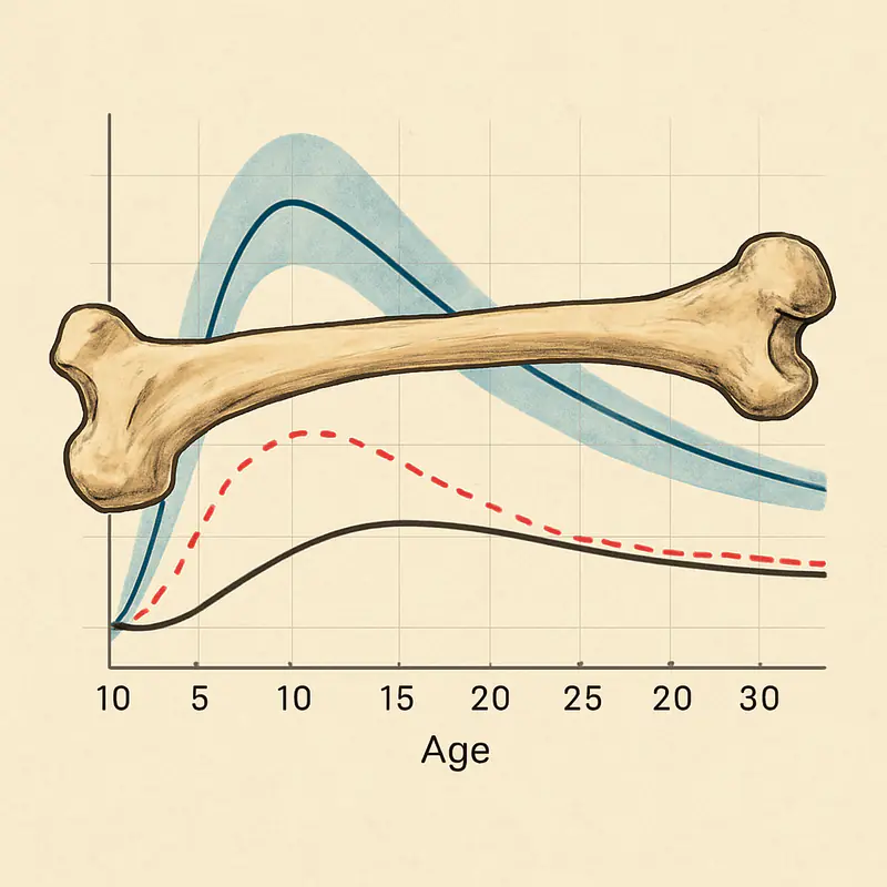
In male subjects, mean clavicular length increased by roughly 34 percent between age 12 and age 25 — approximately 45 millimeters of additional longitudinal growth over that window. In female subjects, the increase over the same period was roughly 26 percent, or approximately 33 millimeters. The slope of growth is not linear. A disproportionate share of that elongation occurs after the typical age at which other long bones have effectively stopped growing — meaning that a seventeen-year-old who assumes his frame is "done" because his height has plateaued is very likely wrong about his clavicle.
Skeletal maturation studies using CT-based classification of the medial clavicular epiphysis put complete fusion of the proximal clavicular physis somewhere between 23 and 27 years in most individuals, with the upper bound extending to approximately 30 years in some cases. Female fusion tends to occur earlier than male fusion, with a measurable acceleration from around age 18 onward in women. Men, on average, retain an open or partially open medial epiphysis significantly longer.
Three practical points follow from those numbers. First, the clavicular growth window is substantially longer than the general growth-plate window people usually cite. Second, the window is wider in men than in women by a meaningful margin. Third, individual variation is large enough that chronological age alone is a poor proxy for biological window status — imaging is the only reliable way to know where a specific individual sits.
Why the Medial Epiphysis Behaves Differently
The medial clavicular physis is unusual in two ways. It is the last to appear — ossification centers don't reliably show up until the mid-teens — and it is the last to fuse. Both of those traits are consequences of the clavicle's embryological origin as an intramembranous bone that later acquires endochondral growth plates at its ends, a hybrid pattern that is not shared by the other long bones of the appendicular skeleton.
For the purposes of a research protocol, what that means is that the tissue responsiveness profile of the medial clavicle in a 24-year-old is more comparable to the distal femur in a 14-year-old than to any other growth plate in a same-age adult. The chondrocytes in the physis are still proliferating. The IGF-1 receptor density is still elevated. The growth factor signaling that drives longitudinal elongation at other sites in early adolescence is still operative here, for a subset of individuals, well into the mid-twenties.
Hormonal Axes and the Research Rationale
The biology of longitudinal bone growth at any physis is ultimately a story about the GH/IGF-1 axis acting on proliferating chondrocytes at the growth plate, mediated by local IGF-1 production in the liver and at the bone itself, and regulated by nutritional status, sleep-stage GH pulses, and mechanical load. Peptides that influence the GH/IGF-1 axis therefore have a mechanistic rationale for influencing clavicular elongation in individuals whose medial epiphysis remains open.
The research-accessible compounds most often discussed in this context fall into three categories. The first is GH secretagogues — growth hormone releasing hormone analogs like CJC-1295 and ghrelin receptor agonists like ipamorelin — which amplify endogenous GH pulses without introducing exogenous GH directly. The second is direct IGF-1 analogs, most notably IGF-1 LR3, whose modified N-terminal extension reduces binding-protein interaction and produces a substantially longer effective half-life than native IGF-1. The third is connective tissue and recovery peptides like BPC-157 and TB-500, which do not act directly on the growth plate but support the training volumes and recovery demands that make mechanical loading protocols sustainable.
None of these compounds have FDA-approved therapeutic indications for skeletal development or frame optimization, and the human data supporting their use for that specific purpose is limited to mechanism-level inference from animal models and adjacent clinical contexts. That is a legitimate gap in the evidence, and any research discussion of the topic should acknowledge it rather than overstate what is known.
The Sourcing Problem
For researchers working in this space, sourcing is a larger concern than is sometimes appreciated. Peptide synthesis at research-grade purity is a nontrivial chemistry problem, and the gap between a product that passes HPLC and mass spectrometry verification and one that does not can be invisible on the outside of the vial. Purity below ~95 percent produces unreliable research outcomes at best and unpredictable off-target effects at worst. Endotoxin contamination from poor manufacturing process control is a secondary concern that rarely shows up on a standard certificate of analysis unless the vendor specifically tests for it.
The practical effect is that vendor selection is a quality-control decision rather than a price decision. Third-party testing documentation, batch-specific certificates of analysis, and transparent disclosure of manufacturing origin are the minimum baseline. Vendors like Pure Tested Peptides have built their reputation specifically on lot-level testing transparency and research-only labeling, which is the kind of documentation chain a serious research protocol should require. Comparable testing depth and catalog breadth across the GH secretagogue and IGF-1 analog categories is available from NuScience Peptides, whose independently verified batch documentation covers the full range of compounds relevant to structural development research.
The point is not brand loyalty. The point is that a researcher who spends months designing a protocol and then sources compounds from an untested supplier has invalidated every other variable in the design.
Mechanical Loading Is Not Optional
The hormonal axis conversation tends to dominate frame-optimization discussions, but the bone remodeling literature is unambiguous on one point: growth factor signaling without mechanical load does not produce targeted structural adaptation. The physis responds to elongation demand. Periosteal apposition — the mechanism by which mature bone increases its cross-sectional area after growth plates have fused — responds to bending and compressive loads transmitted through the shaft.
For the clavicle specifically, the loading movements that transmit the most direct mechanical signal are overhead pressing variants, which apply compressive and bending loads through the acromioclavicular joint, and heavy horizontal pulling, which loads the sternoclavicular and AC joints through different force vectors. Lateral raise variants contribute less direct skeletal loading but significantly influence the muscular frame that visually amplifies clavicular width. Progressive overload on these movements across training cycles of months rather than weeks is the mechanical substrate that any peptide protocol is supposed to compound, not replace.
Window Assessment Before Protocol Design
The single most useful piece of information a researcher in this area can have is the current ossification status of their own medial clavicular epiphysis. CT-based classification schemes — most prominently the Schmeling-Kellinghaus five-stage system — map the progression from unfused through active fusion to complete ossification of the physeal line. An individual in stage 2 or early stage 3 has substantially different biological potential from one in stage 4 or 5, and the difference is not inferable from chronological age alone.
For the 22- to 28-year-old cohort that dominates frame-optimization discussions, imaging assessment is the only way to know whether a research protocol targeting the physis has a biological substrate to act on at all. Protocols designed without that information are operating on statistical averages that may not apply to the specific individual running them.
The Honest Summary
The clavicle continues to grow meaningfully into the mid-twenties in a substantial fraction of healthy individuals, with male subjects retaining biological potential later than female subjects, and with individual variation wide enough that population averages should not be treated as personal deadlines. The mechanistic case for peptide research targeting the GH/IGF-1 axis during this window is grounded in established bone biology, but the direct human evidence for the specific outcome of clavicular elongation remains thin. Mechanical loading is the signal the hormonal environment is supposed to amplify, and sourcing quality is the variable that determines whether any of the research is interpretable at all.
Frame optimization is not a magic trick. It is a set of overlapping biological inputs — hormonal, mechanical, nutritional, temporal — applied to a specific skeletal structure during a specific biological window that, for the clavicle, lasts longer than most people assume.